Home /
Expert Answers /
Biology /
attached-is-a-planaria-slide-under-the-microscope-at-the-low-medium-and-high-power-objectives-the-pa552
(Solved): Attached is a Planaria slide under the microscope at the low, medium and high power objectives. The ...
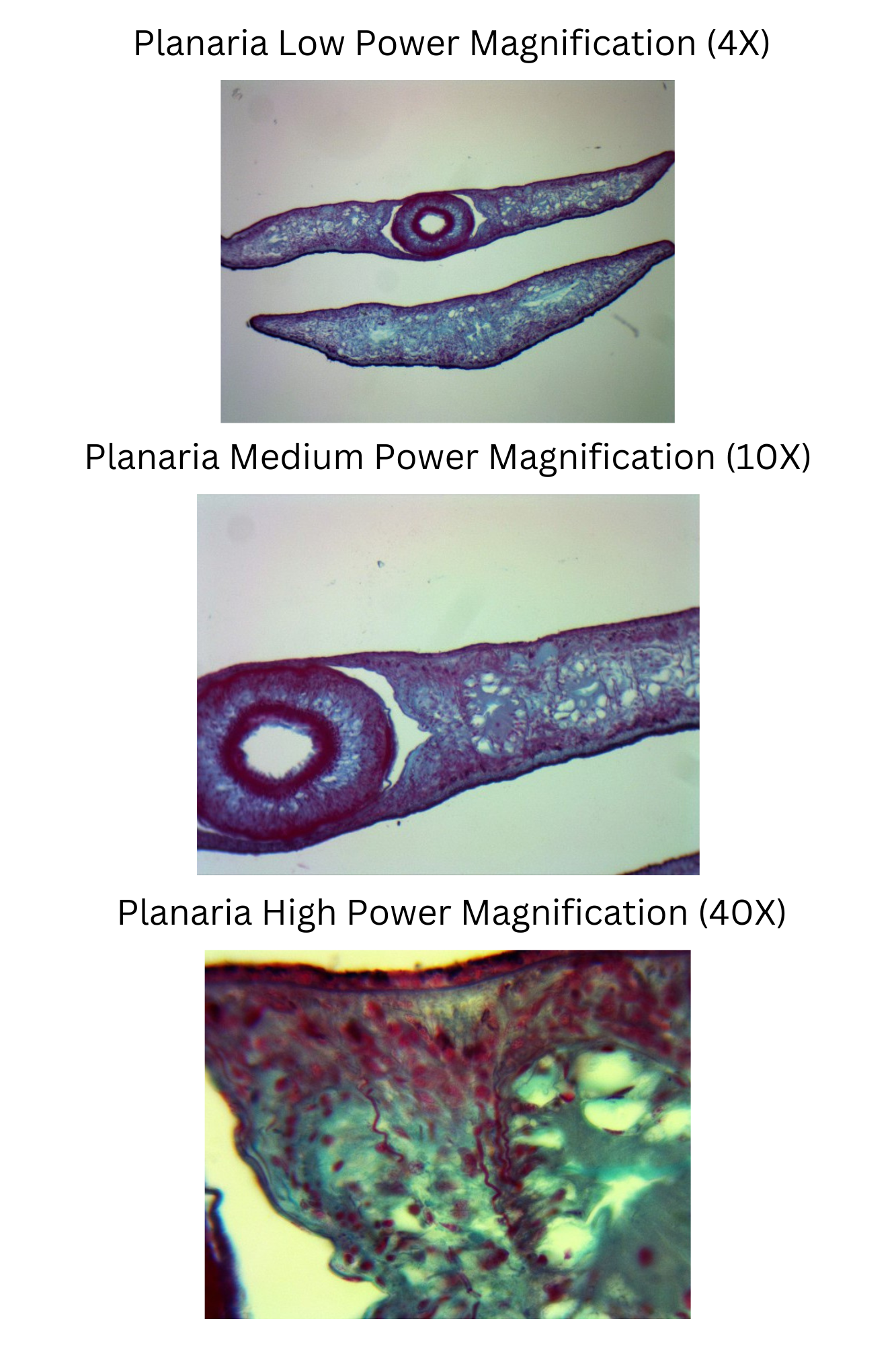
Attached is a Planaria slide under the microscope at the low, medium and high power objectives. The magnifications should locate and focus on the two cross sections of the Planaria specimen. Choose the magnification that best displays the pharynx, intestine, and ciliated epidermis. Record the total magnification of the microscope lenses used for the photo. Label the pharynx, intestine, and ciliated epidermis on the chosen magnification. Note: If you cannot identify a structure, indicate the reason as a comment. Planaria Low Power Magnification (4X) Planaria Medium Power Magnification (10X) Planaria High Power Magnification (40X)